Hydronephrosis
Swelling of the kidney, often found on a pregnancy scan.
What is Hydronephrosis?
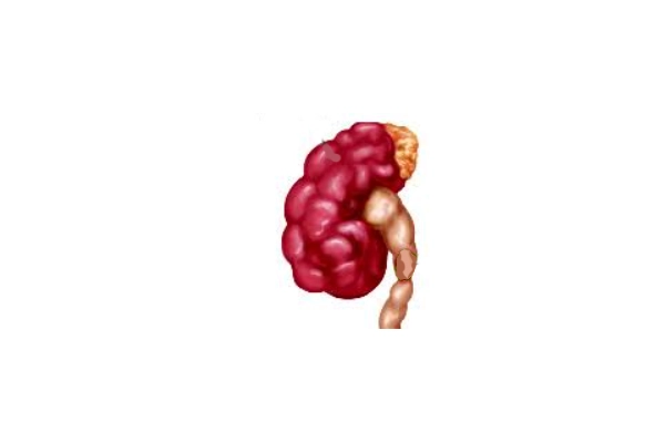
Hydronephrosis means the collecting system of the kidney is swollen with urine. It is a finding, not a diagnosis in itself — the real question is always why.
It is most often picked up on a routine scan during pregnancy. Parents are told the baby’s kidney is swollen, and understandably become very anxious. In reality a large proportion of these babies have mild swelling that settles by itself in the first year or two.
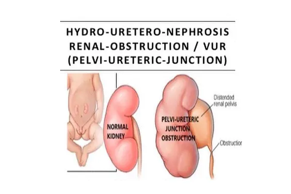
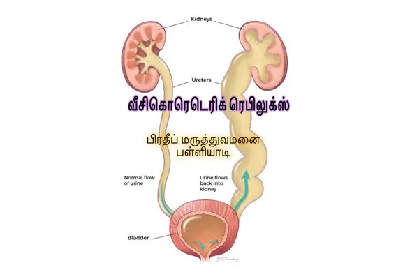
The causes that matter are blockage at the junction of the kidney and the ureter (PUJ obstruction), blockage where the ureter meets the bladder, urine flowing backwards (reflux), and in boys, posterior urethral valves.
The purpose of the tests after birth is to separate the babies whose kidney is simply roomy from the few whose kidney is genuinely obstructed and losing function.
In your language
Hydronephrosis
Swelling of the kidney, often found on a pregnancy scan.
Hydronephrosis means the collecting system of the kidney is swollen with urine. It is a finding, not a diagnosis in itself — the real question is always why.
சிறுநீரகத்தில் நீர் தேங்குதல்
கர்ப்ப கால ஸ்கேனில் “குழந்தையின் சிறுநீரகத்தில் நீர் தேங்கியுள்ளது” என்று சொன்னால் பயப்பட வேண்டாம். பெரும்பாலான குழந்தைகளுக்கு இது லேசானது; ஓராண்டுக்குள் தானாகவே சரியாகிவிடும். பிறந்த பிறகு ஸ்கேன் மற்றும் தேவைப்பட்டால் MCUG, ரீனோகிராம் பரிசோதனைகள் மூலம் உண்மையான அடைப்பு உள்ளதா என்று கண்டறியப்படும். அடைப்பு இருந்து சிறுநீரகத்தின் செயல்திறன் குறைந்தால் மட்டுமே அறுவை சிகிச்சை தேவை. பரிசோதனைகளைத் தவிர்க்காமல் தொடர் கண்காணிப்பு அவசியம்.
വൃക്കയിൽ വെള്ളം കെട്ടിനിൽക്കുന്ന അവസ്ഥ
ഗർഭകാല സ്കാനിൽ “കുഞ്ഞിന്റെ വൃക്കയിൽ വെള്ളം കെട്ടിനിൽക്കുന്നു” എന്ന് പറഞ്ഞാൽ പരിഭ്രമിക്കേണ്ട. മിക്ക കുഞ്ഞുങ്ങളിലും ഇത് നിസ്സാരമാണ്, ഒന്നു രണ്ടു വർഷത്തിനുള്ളിൽ സ്വയം മാറും. ജനനശേഷം സ്കാൻ, ആവശ്യമെങ്കിൽ MCUG, റീനോഗ്രാം എന്നിവയിലൂടെ യഥാർത്ഥ തടസ്സമുണ്ടോ എന്ന് കണ്ടെത്തും. തടസ്സം മൂലം വൃക്കയുടെ പ്രവർത്തനം കുറയുമ്പോൾ മാത്രമേ ശസ്ത്രക്രിയ വേണ്ടിവരൂ. തുടർ പരിശോധനകൾ ഒഴിവാക്കരുത്.
What Parents Notice
Not every child shows every sign. If something on this list matches your child, it is worth an examination rather than a guess.
- Very often none — the swelling is seen on a scan before birth and the baby appears completely well
- Repeated urinary tract infections or unexplained fever
- A lump felt in the tummy in a baby
- Episodes of tummy or flank pain, sometimes with vomiting, in an older child
- Poor weight gain or feeding difficulty in a newborn
- In boys with valves: a poor urinary stream, straining, or dribbling
How It Is Confirmed
- Ultrasound after birth — usually repeated after the first 48 hours to 7 days, because a newborn is relatively dry in the first days and a scan done too early can underestimate the swelling
- MCUG to look for reflux or posterior urethral valves, particularly in boys and in more marked swelling
- Diuretic renogram (DTPA or EC scan) — the key test for obstruction: it shows how much each kidney contributes and whether urine drains freely
- Blood tests for kidney function where both kidneys are affected
- Serial ultrasound follow-up, because the pattern over time is more informative than any single scan
How It Is Treated
- Mild hydronephrosis: observation with repeat ultrasound. Most improves or resolves without any intervention.
- Moderate hydronephrosis: surveillance scans and renograms, often with preventive antibiotics in infancy.
- Obstruction at the PUJ with poor drainage or falling function: pyeloplasty, an operation that removes the narrow segment and rejoins the kidney to the ureter. It can be done open or laparoscopically.
- Reflux: managed as for vesicoureteral reflux.
- Posterior urethral valves in a boy: the valves are removed endoscopically, promptly.
- Infection, when it occurs, is treated fully — a swollen kidney with infection is a more urgent situation than a swollen kidney alone.
Does My Child Need Surgery?
Not every child with Hydronephrosis needs an operation. These are the situations in which surgery is advised — and the decision is always made after examining your child, never from a description alone.
The renogram shows genuine obstruction with impaired drainage
The affected kidney’s function falls on repeat testing
The swelling increases progressively on serial ultrasound
The child has pain, a palpable lump, or repeated kidney infections
There is a treatable cause such as posterior urethral valves, which is dealt with without waiting
What Happens Over Time
Most swelling picked up before birth is mild and settles on its own within the first year or two. Where an obstruction is found and relieved in time, drainage improves and the kidney keeps the function it had. The purpose of follow-up is to catch the small number of kidneys that are quietly losing function.
This describes what usually happens, not what will happen to your child. The surgeon will give you a specific picture after examining them.
Can It Be Prevented?
It cannot be prevented, because it develops before birth. What can be prevented is damage from delay, by attending the scans arranged after birth.
From First Visit to Going Home
What happens, in order, so that nothing on the day comes as a surprise.
Consultation
Dr. Pratheep Samraj examines your child and goes through the history, earlier reports and any treatment already given.
Investigation
Only the tests that change the decision are asked for — commonly an ultrasound, blood tests, and occasionally an X-ray or a scopy.
Explanation
You are told what the condition is, whether it needs surgery, what happens if you wait, and what the operation involves.
Treatment
If surgery is appropriate it is planned with paediatric anaesthetic cover, with ICU and NICU support available in the hospital.
Follow-up
You go home with written instructions and a review date, and you can reach the hospital by phone or WhatsApp in between.
Hospital stay
Observation needs no admission. Pyeloplasty usually means 3 to 5 days in hospital; endoscopic valve treatment is shorter.
Recovery at home
After pyeloplasty a drainage stent may stay for a few weeks and is removed in a short second procedure. Normal activity within 2 to 3 weeks.
Follow-up
Long-term. Ultrasound and, where needed, renograms continue for years — the kidney is watched as the child grows.
Please bring with you: all previous scan reports, blood test reports, discharge summaries, records of any earlier operation, current medicines, and your child’s immunisation card. Photographs on your phone of a swelling that comes and goes are genuinely useful.
Please note: this page is general information about Hydronephrosis, not advice about your child. Timing, suitability for surgery and the expected result differ from child to child and are decided only after clinical evaluation.
Questions Parents Ask
If your question is not here, send it on WhatsApp — you will get an answer from the hospital team.

Dr. Pratheep Samraj
Consultant Paediatric & Neonatal Surgeon · Palliyadi, Kanyakumari
Super-specialist surgical care for newborns, infants and children, with expertise in paediatric urology, congenital anomalies, gastro-intestinal surgery and minimally invasive surgery.
Related Conditions
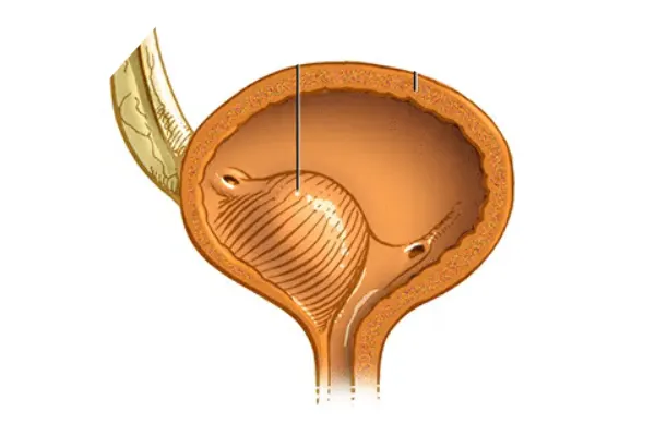
A kidney seen swollen on the pregnancy scan needs following up, not panicking about.
Most of it settles by itself; the point of follow-up is to catch the minority that is a genuine blockage before function is lost. Bring the antenatal scan reports with you.
Consultation 10:00 AM - 1:00 PM & 4:00 PM - 8:00 PM · 24-hour laboratory & pharmacy
In an emergency — green vomiting in a baby, a groin swelling that will not go back, severe tummy pain, or a newborn not passing urine or meconium — come straight to the hospital or to your nearest emergency department. Do not wait for a reply to a message.